class: center, middle, inverse, title-slide .title[ # TDA to characterize spatial distributions ] .subtitle[ ## Looking at mRNA at sub-cellular resolutions ] .author[ ### <strong>Erik Amézquita</strong>, Sutton Tennant, Sandra Thibivillers, Sai Subhash<br>Benjamin Smith, Samik Bhattacharya, Jasper Kläver, Marc Libault<br>—<br>Division of Plant Science & Technology<br>Department of Mathematics<br>University of Missouri<br>— ] .date[ ### 2025-05-21 ] --- background-image: url("../../demat/figs/fam9_3.png") background-size: 100px background-position: 98% 2% # About me: From MX to MI to MO at MU ## I work across multiple disciplines and countries .left-column[  ] .right-column[ - 2013 - 2018 : Licenciatura (Bachelor): Mathematics @ Universidad de Guanajuato and CIMAT. Thesis focused on Topological Data Analysis applied to archaeology. - 2018 - 2023 : PhD: Computational Mathematics, Science, and Engineering @ Michigan State University. Dissertation: Exploring the mathematical shape of plants. **Came for the math. Stayed for the plants.** - 2023 - 2025 : ~~PFFFD~~ ~~PFFIE~~ PFF Postdoctoral Fellow @ Division of Plant Science & Technology (80%) / Department of Mathematics (20%) at MU. - Sept. 2025 : Assistant Professor at MU. Division of Plant Science & Technology. ] <br><br><br><br><br><br><br><br><br><br><br><br><br><br> --- # Phenotyping the shape of things to come <div class="row" style="font-family: 'Yanone Kaffeesatz'; font-size:22px;"> <div class="column" style="max-width:33%"> <p style="line-height:0;text-align: center; font-size:28px">Phenotyping patterns</p> <img style="padding: 0 30px;" src="../figs/molecular_cartography_2x2.jpg"></img> <img style="padding: 0 30px;" src="../figs/persistence_images_1x1.svg"></img> <p style="text-align: center;">mRNA sub-cellular localization in soybean nodule cells.</p> </div> <div class="column" style="max-width:33%"> <p style="line-height:0;font-size:28px;text-align: center;">Phenotyping movement</p> <img style="padding: 0 10px;" src="../../cuscuta/figs/qmlphv.gif"></img> <img style="padding: 35px 30px;" src="../../cuscuta/figs/avg_pl.svg"></img> <p style="text-align: center;">Tracking and describing <i>Cuscuta campestris</i> circumnutation</p> </div> <div class="column" style="max-width:33%"> <p style="line-height:0;font-size:28px;text-align: center;">Phenotyping data</p> <img style="padding: 0 0px;" src="../../nasrin/figs/fpkm_raw_3.png"></img> <img style="padding: 30px 0px;" src="../../nasrin/figs/lung_fpkm_meancorr_eps1.0e+06_r40_g40.png"></img> <p style="text-align: center;">Reducing <strong>and</strong> clustering high-dimensional omics data</p> </div> </div> --- background-image: url("../../barley/figs/seed.png") background-size: 325px background-position: 99% 99% class: middle # Roadmap for today 1. Biological context: genomics analysis to characterize cell type - Central dogma - mRNA localization 1. TDA to model mRNA localization patterns - Persistence images 1. Results - Modeling sub-cellular mRNA localizations --- class: inverse, middle, center # 1. A crash course in genomics analysis ## Characterizing cell types --- # The Central Dogma and Gene Expression .pull-left[  <p style="font-size: 10px; text-align: right; color: Grey;">Credits: <a href="https://doi.org/10.1242/bio.017178">Xu <em>et al.</em> (2016)</a></p> ] .pull-right[ - Every cell in an organism has the exact same* DNA. - But cells will only express a subset of genes depending on their type. - By knowing which genes are overexpressed, we can gather what makes each cell type unique and what are their exact functions.  ] --- # mRNA localization at a sub-cellular level - Beyond gene expression counts: Spatial segregation and asymmetrical distribution of mRNA across the cytosol in the soybean nodule. - Molecular Cartography™ data provided by the Libault Lab .pull-left[  Infected soybean nodule cells. Glyma.05G092200 in green. Glyma.05G216000 in red. ] .pull-right[ **Goals**: "How patterny is a pattern?" - Quantify the spatial patterns followed by mRNA within individual cells. - Mathematically model all observed mRNA sub-cellular distributions. - *Use this mathematical model to differentiate cell types and genotypes.* **Challenge** - Develop a mathematical model that works for any cell size, orientation, shape, and dimension. ] --- # Traditional model: Density of transcripts   But this characterization discards sub-cellular information! --- # Same density, different patterns  - 97 genes (including 10 bacterial ones) → 2 genes - 2938 cells → 918 infected ones. **Subcellular transcript patterns ↔ spatial location of the cell within the nodule** --- class: inverse, middle, center # Alternate model: Topological Data Analysis 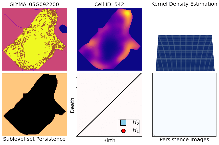 --- class: inverse, middle, center # 2. Characterizing mRNA spatial patterns and distributions ## with Topological Data Analysis (TDA) --- # Alternate model: Topological Data Analysis 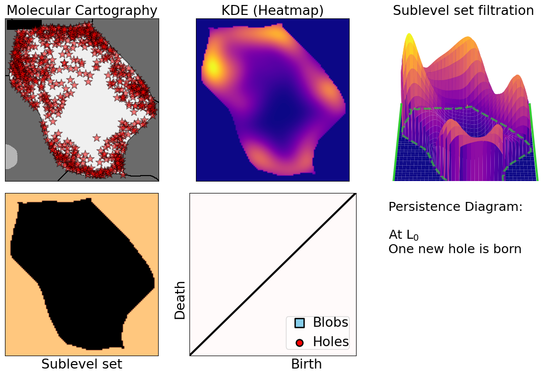 --- # Alternate model: Topological Data Analysis  --- # Alternate model: Topological Data Analysis 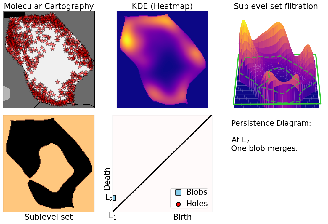 --- # Alternate model: Topological Data Analysis 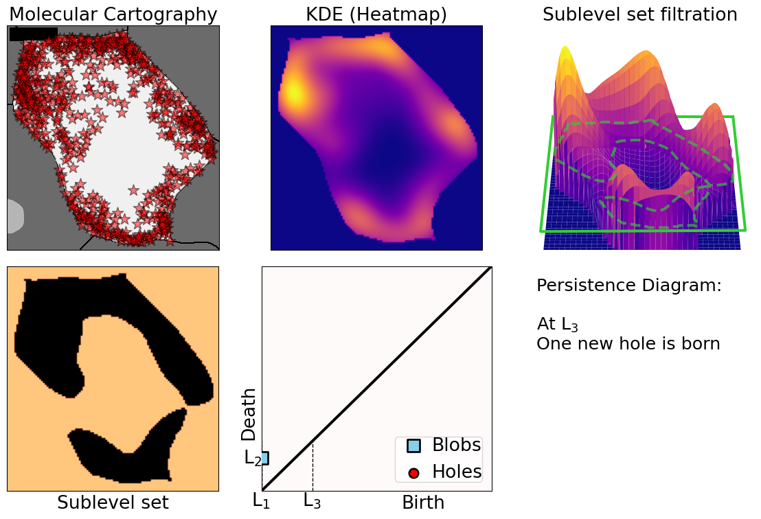 --- # Alternate model: Topological Data Analysis  --- # Alternate model: Topological Data Analysis  --- # Alternate model: Topological Data Analysis  --- # Alternate model: Topological Data Analysis  --- # Alternate model: Topological Data Analysis  --- # Alternate model: Topological Data Analysis  --- # Alternate model: Topological Data Analysis  --- # Alternate model: Topological Data Analysis  --- # Mathematical motivation: stability - [**Theorem (Cohen-Steiner, Edelsbrunner, Harer, 2007)**](https://doi.org/10.1007/s00454-006-1276-5): Given a nice enough topological space `\(\mathbb{X}\)` and two nice enough filtration functions `\(f,g:\mathbb{X}\to\mathbb{R}\)`, then `$$d_B(\text{dgm}(f), \text{dgm}(g)) \leq \|f-g\|_\infty,$$` - where `\(d_B\)` is the bottleneck distance. - **Translation**: If the original complex wiggles a tiny bit, then the elements of its related persistence diagram will wiggle only a tiny bit as well. ## However - Outside stable distances, it is hard to do anything interesting in the space of persistence diagrams. - E.g.: there are no unique means! - Hard to perform Machine Learning directly with persistence diagrams --- # Rotate 45 degrees for ML ammenability  --- # TDA: From patterns to numbers  --- # Mathematical justifications - **Definition:** Given two persistence diagrams `\(D_1, D_2\)`, for `\(1\leq p<\infty\)`, we define the *p-Wasserstein* distance between them as `$$W_p(D_1, D_2) := \inf_{\gamma:D_1\to D_2}\left(\sum_{u\in D_1} \left\| u-\gamma(u) \right\|_\infty^p\right)^{1/p},$$` where the infimum is over all possible bijections `\(\gamma: D_1\to D_2\)`. - **Theorem [[Mileyko *et al* (2011)](https://doi.org/10.1088/0266-5611/27/12/124007)]:** For nice filtrations, the persistence diagrams are Wasserstein-stable under small perturbations of the data they summarize. - **Theorem [[Adams *et al.* (2017)](http://jmlr.org/papers/v18/16-337.html)]:** The persistence image `\(I(D)\)` of a persistence diagram `\(D\)` with Gaussian distributions is stable with respect to the 1-Wasserstein distance between diagrams. ### If the overall shape/pattern is perturbed a little bit, then the resulting persistence images are perturbed only a little bit as well --- class: inverse, middle, center # 3. Results ## Do TDA for all cell-gene combinations ### One gene at a time vs multiple genes --- # Focus on `\(H_1\)` and `\(H_2\)` <img src="../figs/molecular_cartography_2x4.png" width="500" style="display: block; margin: auto;" /> <img src="../figs/persistence_images_2x4.png" width="600" style="display: block; margin: auto;" /> --- background-image: url("../figs/bw25_scale32_-_PI_1_1_1_H1+2_cell_sample.png") background-size: 620px background-position: 75% 99% # PCA on all topological descriptors <img src="../figs/bw25_both_scale16_-_PI_1_1_1_pca_H1+2_gridded.png" width="350" style="display: block; margin: auto auto auto 0;" /> --- background-image: url("../figs/bw25_scale32_-_PI_1_1_1_H1+2_kde_sample.png") background-size: 620px background-position: 75% 99% # Show me <img src="../figs/bw25_both_scale16_-_PI_1_1_1_pca_H1+2_gridded.png" width="350" style="display: block; margin: auto auto auto 0;" /> --- background-image: url("../figs/bw25_scale32_-_PI_1_1_1_H1+2_kde_sample.png") background-size: 620px background-position: 99% 50% # Show me .left-column[ ### PC 1 - Related to the number of distinct hotspots - Correlated to transcript number and cell size ### PC 2 - Related to the heterogeneity of hotspots - Correlated to transcript density ] --- background-image: url("../figs/GLYMA_05G216000_bw15_PI_scale8_kde_sample.png") background-size: 800px background-position: 50% 75% # Similar observations with other genes --- background-image: url("../figs/GLYMA_12G027400_bw20_PI_scale40_kde_sample.png") background-size: 600px background-position: 50% 75% # Similar observations with other genes --- background-image: url("../figs/GLYMA_15G210100_bw15_PI_scale48_kde_sample.png") background-size: 750px background-position: 50% 95% # Similar observations with other genes --- background-image: url("../figs/GLYMA_17G185800_bw15_PI_scale24_kde_sample.png") background-size: 650px background-position: 50% 99% # Similar observations with other genes --- # Connecting PC 02 to the biological context  - Senescent cells exhibit a distinct transcriptomic spatial pattern compared to the rest of population. - Loss of mRNA localization may be a lesser known contributor to cell senescence. --- # Connecting PC 02 to the biological context  - Senescent cells exhibit a distinct transcriptomic spatial pattern compared to the rest of population. - Loss of mRNA localization may be a lesser known contributor to cell senescence. --- # We define a morphospace of transcriptomic patterns  # We then work "backward" --- class: bottom background-image: url("../figs/scale32_-_PI_1_1_1_H1+2_synthetic_30_clusters.jpg") background-size: 900px background-position: 50% 1% <img src="../figs/scale32_-_PI_1_1_1_H1+2_synthetic_pca_30_clusters.jpg" width="600" style="display: block; margin: auto;" /> --- class: bottom background-image: url("../figs/scale32_-_PI_1_1_1_H1+2_synthetic_varclusters.jpg") background-size: 900px background-position: 50% 1% <img src="../figs/scale32_-_PI_1_1_1_H1+2_synthetic_pca_varclusters.jpg" width="600" style="display: block; margin: auto;" /> --- # Discussion and future directions **Biologically speaking** - Senescent cells exhibit a distinct transcriptomic spatial pattern compared to the rest of population. - Loss of mRNA localization may be a lesser known contributor to cell senescence. - *How does the morphospace of patterns change if we take into account more genes, more cell types, more tissues, and more mutants?* **Mathematically speaking** - Topological Data Analysis offers a robust way to encode the shape of patterns. - Robust to differences in scale, underlying boundaries, or orientation. - The framework is open to any number of cells, genes, and dimensions. <img src="../figs/D2_GLYMA_05G092200_z_kde_pd_suplevel_by_both_00512.jpg" width="550" style="display: block; margin: auto;" /> --- # Software used .pull-left[ - All the work has been done in python with mostly standard libries (`numpy`, `scikit-learn`, `matplotlib`, `pandas`, etc.) - KDEs computed efficiently with [`KDEpy`](https://kdepy.readthedocs.io/en/latest/) - Sublevel set filtration of images and persistence diagrams done with [`gudhi`](https://gudhi.inria.fr/)  ] .pull-right[ - Persistence Images computed with [`persim`](https://persim.scikit-tda.org/en/latest/)  ] --- class: inverse # Thank you! <div class="row"> <div class="column" style="max-width:60%; font-size: 15px;"> <img style="padding: 0 0 0 0;" src="https://bondlsc.missouri.edu/wp-content/uploads/2023/11/libault-returns-to-bond-lsc-as-plant-scientist-with-single-cell-focus-1.jpg"> </div> <div class="column" style="max-width:40%; font-size: 24px; line-height:1.25"> <p style="text-align: center;"><strong>Contact and slides:</strong></p> <p style="text-align: center;color:Blue">eah4d@missouri.edu</p> <p style="text-align: center;color:Blue">ejamezquita.github.io</p> </div> </div> <div class="row"> <div class="column" style="max-width:35%; font-size: 20px;"> <p style="font-size: 25px; text-align: center;"> Libault Lab (MU) </p> <ul> <li><strong>Marc Libault</strong></li> <li><strong>Sandra Thibivillers</strong></li> <li>Hengping Xu</li> <li>Sahand Amini</li> <li>Hong Fu</li> <li><strong>Sutton Tennant</strong></li> <li>Md Sabbir Hossain</li> <ul> </div> <div class="column" style="max-width:65%; font-size: 20px;"> <p style="font-size: 25px; text-align: center;"> With help from:</p> <ul> <li>Sai Subash (Nebraska-Lincoln)</li> <li>Benjamin Smith (UC Berkeley)</li> <li>Samik Bhattacharya (Resolve Biosciences)</li> <li>Jasper Kläver (Resolve Biosciences)</li> <ul> </div> </div>